Figure 4.

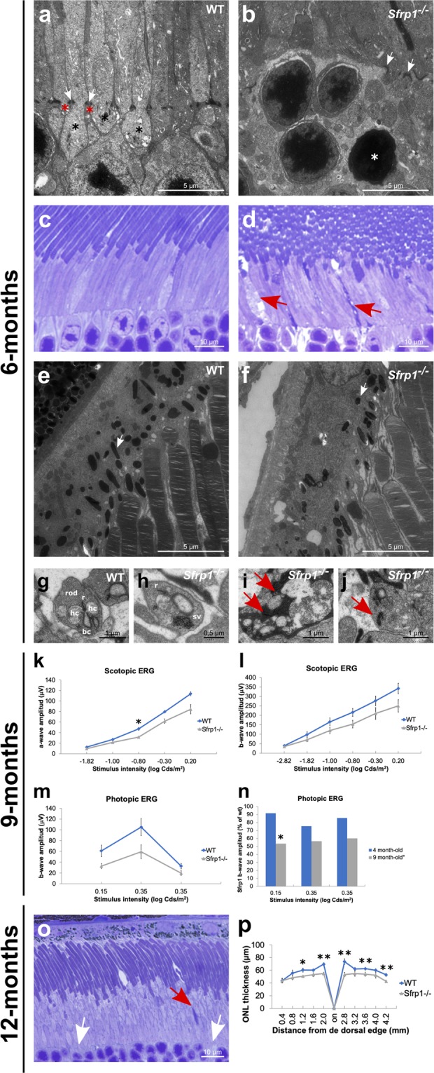
OLM is disrupted in the retina of Sfrp1−/− mice in association with an impoverishment of visual function. (a,b; e–j) TEM analysis of the retina from 6 months-old wt and Sfrp1−/− mice. Images show the OLM (a,b), RPE (e,f) and photoreceptor-bipolar cells synapses (g–j). White arrows in a,b point to adherens junctions between Müller glial cell end-feet (red asterisks) and photoreceptors (black asterisks). White arrows in e,f point to melanosomes. Red arrows in i,j indicate synaptic terminal disorganization. (c,d) Semi-thin frontal sections from 6 months-old wt and Sfrp1−/− retinas stained with toluidine blue. Note the abnormal presence of degenerating outer segments in the mutant retina (red arrows in d). (k,l) The graphs show the ERG recordings of rods and rod bipolar cells (scotopic, k,l) and cone –pathway (photopic, m) response to light of 9 months-old wt and Sfrp1−/− animals. (n) The graph represents the activity of photoreceptors from 4 and 9 months-old Sfrp1−/− mice. Note that activity is expressed as a percentage of wt age-matched animals. Only 9 months-old Sfrp1 mice showed a significantly reduced activity relative to their control. (o) Semi-thin frontal sections from 10 months-old Sfrp1−/− retina stained with toluidine blue. Note the continuous presence of degenerating (red arrows) and enlarged OS with evident breakage of the OLM (white arrows); (p) The graphs represent the thickness of the ONL in 12 months-old wt and Sfrp1−/− retinas showing a significant reduction in the mutants more evident in central regions. Mann-Whitney U test; *p < 0.05, **p < 0.01. Abbreviations. bc: bipolar cell; hc: horizontal cell; r ribbon; sv: synaptic vesicles.
