Figure 6.

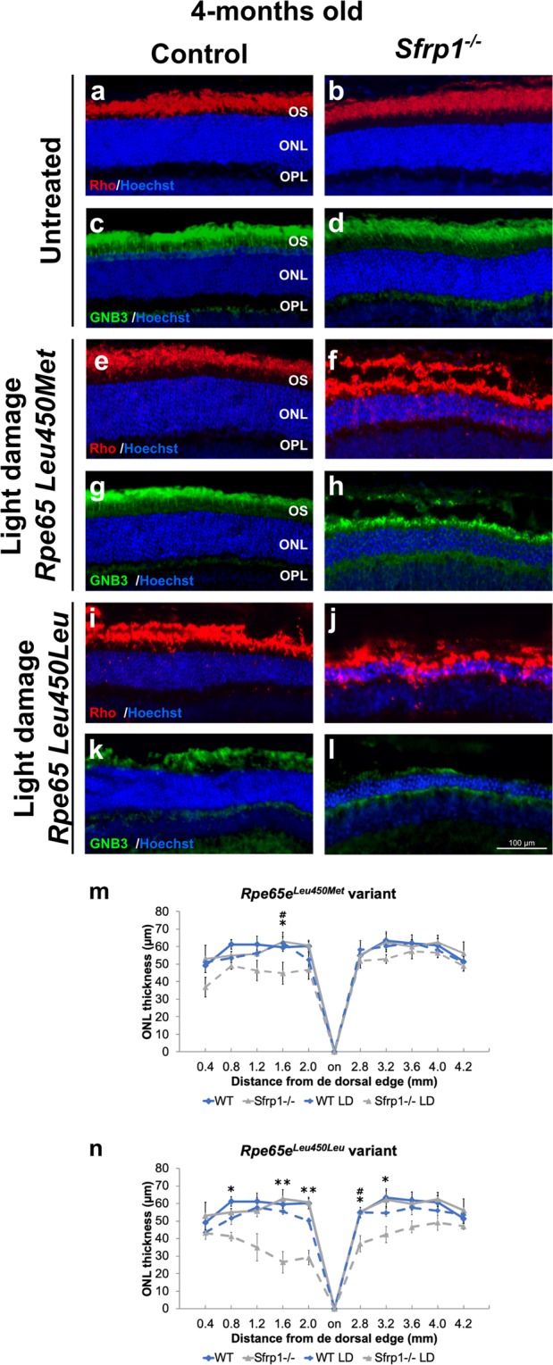
Sfrp1−/− retinas are more prone to light-induced damage even in the presence of the Rpe65Leu450Met protective variant. (a–l) Frontal cryostat sections from 4 months-old control and Sfrp1−/− retinas carrying the Rpe65Leu450Leu or the protective Rpe65eLeu450Met variants before and 7 days after light induced-damage (light conditions: 15000 lux of cool white fluorescent light for 8 hrs). Sections were immunostained with antibodies against Rho (rods, red) or GNB3 (cones, green) and counterstained with Hoechst. (m,n) The graphs represent the ONL thickness (measures were taken in cryostat frontal sections of the eye at the level of the optic disk) in the different conditions. Kruskal Wallis with post hoc Dunn. *Indicates significant differences between Sfrp1−/− untreated and light-damage mice. #Indicates significant difference between Sfrp1−/− and wt animals exposed to light damage. No significant differences were found between wt untreated and light-damage mice. *p < 0.05, **p < 0.01. Scale bar in l applies to all panels. Abbreviations: ONL: outer nuclear layer; OPL: outer plexiform layer; OS outer segment.
