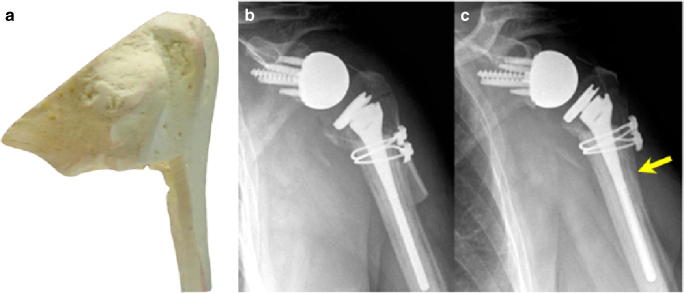
Fig. 3.
Images adapted from Chacon et al. A Sawbones model of the prepared proximal humeral allograft (a). Immediate post-operative (b) and last available radiographs (c) demonstrating incorporation at the allograft-bone junction in both the metaphyseal region and the diaphyseal region (arrow) [40]

