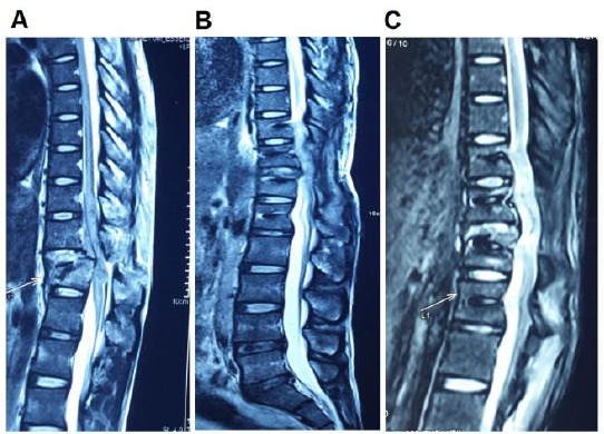
Figure 5.

Comparison of a lesion in MRI; A) Before treatment; B) 3 months after the first treatment; and C) 6 months after the first treatment; The MRI image of the lesion region was taken before the treatment A); 3 months after the first treatment B); and 6 months after the first treatment C); B) and C) present the recovery of the injured spinal cord
