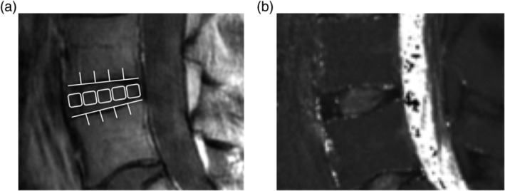
Figure 2.

In second echo image, disc was divided into five areas, designating the front of the anterior annulus fibrosus (AF), the middle of the nucleus pulposus (NP), and the last of the posterior AF (A). In the same region, we measured the mean values (B)
