Fig. 5.
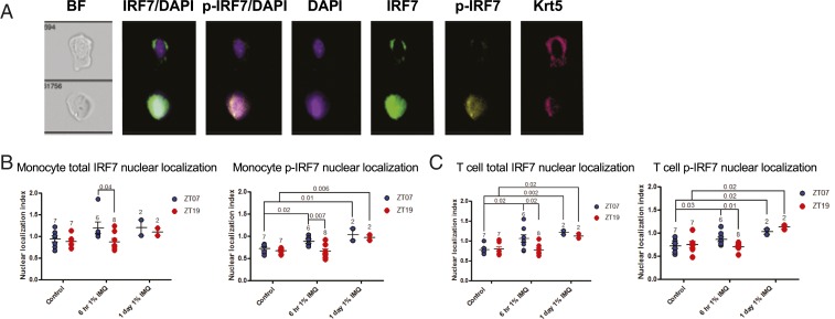
IMQ-induced nuclear localization of total and phosphorylated IRF7 is diurnal in epidermal T cells and monocytes. Male Wt mice were treated with 1% IMQ for 6 h or 1 d during the day (ZT07) or night (ZT19) and back epidermis was dissociated, stained for cell surface markers and intracellular IRF7 and p-IRF7, and analyzed on the ImageStream Flow Cytometer. Nuclear localization index was calculated using IDEAS software. (A) Example images (60x) taken during ImageStream fluorescence imaging, showing a cell with no p-IRF7 nuclear localization (Top) and one with p-IRF7 nuclear localization (Bottom). (B) Average nuclear translocation index for total IRF7 (Left) and p-IRF7 (Right) in monocytes (CD45+CD11c−CD3e−). (C) Average nuclear translocation index for total IRF7 (Left) and p-IRF7 (Right) in T cells (CD45+CD3e+CD11c−). (A–C) Each data point represents one mouse, and mean ± SEM is indicated. Statistical significance was determined by Student’s paired t test and significant or near-significant P values are shown.

