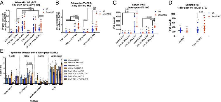
Fig. 6.
Systemic Bmal1 deletion results in exacerbated IMQ-induced serum IFN-β and ISG expression in the skin. (A) Irf7 qPCR on whole back skin from Wt (blue, n = 17 to 30) and Bmal1 KO mice (red, n = 6 to 9) after 6 h or 1 d of 1% IMQ during the day (ZT07) or night (ZT19). (B) Irf7 qPCR on isolated epidermis from Wt (blue, n = 10 to 18) and Bmal1 KO mice (red, n = 4 to 8) after 1% IMQ during the day (ZT07) or night (ZT19) for 1 d. (C) Wt serum IFN-β after 2 or 6 h of 1% IMQ during the day (ZT07) or night (ZT19). (D) Wt (blue) vs. Bmal1 KO (red) serum IFN-β after 1 d of 1% IMQ at ZT07. (E) Flow cytometry quantification of immune cell populations within the epidermis in Wt and Bmal1 KO mice treated with 1% IMQ for 6 h during the day or night (indicated by white and black bars). (A–E) Each data point represents one mouse, and mean ± SEM is indicated. The numbers above each group indicate the number of samples analyzed. Statistical significance was determined by Student’s paired t test and significant or near-significant P values are shown.

