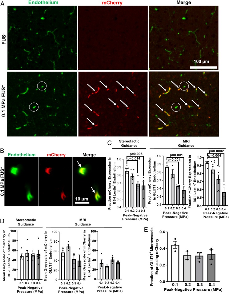
Fig. 1.
FUS peak-negative acoustic pressure (PNP) may be tuned to yield sonoselective cerebrovascular endothelial transfection. (A and B) Confocal images of FUS+ (0.1 MPa) and contralateral FUS− brain tissue showing expression of mCherry reporter gene (red) with respect to endothelial cells (BS-I lectin, green). Arrows denote mCherry colocalization with endothelium. Circles denote untransfected capillaries. (C) Bar graphs of fraction of mCherry expression in cerebrovascular endothelium as a function of PNP. Highly selective endothelial transfection is observed at low PNPs (i.e., 0.1 MPa and 0.2 MPa). Similar relationships were observed when using both stereotactic and MR image guidance and both GLUT1 and BS-I lectin as endothelial markers. One-way ANOVAs followed by Dunnett’s multiple comparison tests. (D) Bar graphs of mean grayscale intensity of mCherry transgene expression in endothelium. Increasing PNP did not enhance endothelial mCherry fluorescence intensity. One-way ANOVAs followed by Dunnett’s multiple comparison tests. (E) Bar graph of fraction of GLUT1+ microvessels expressing mCherry. Increasing PNP did not increase the fraction of transfected microvessels. One-way ANOVA followed by Dunnett’s multiple comparison tests.

