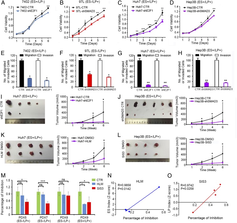
Fig. 5.
Targeting oncofetal drivers might be a novel effective strategy in HCC treatment. (A) HCC cells with ES-like signature were stably transfected with shRNAs specifically targeting E2F1 (shE2F1) or control shRNAs (CTR). The cell growth rates were determined using an MTT assay. (B) HCC cells with LP-like signature were stably transfected with shRNAs specifically targeting SMAD3 (shSMAD3) or control shRNAs (CTR). The cell growth rates were determined using an MTT assay. (C and D) HCC cells with mixed signature were stably transfected with shE2F1 or shSMAD3, and the cell growth rates were determined using an MTT assay. (E–H) Cells were seeded in the cell migration or invasion chamber at a density of 5,000 cells per well. The migrated or invaded cells were counted 48 h later. (I) Huh7-CTR or Huh7-shE2F1 cells were subcutaneously injected into the left dorsal and right dorsal flank of nude mice. The tumor volumes were monitored for 6 wk. (J) Xenograft tumor assays were also performed under similar conditions in Hep3B-CTR and Hep3B-shSMAD3 cells. (K) Huh7 xenograft tumors were implanted in to the left dorsal flank of nude mice, and E2F1 inhibitor HLM6474 (20 mg/kg) were intraperitoneally injected into the tumor-bearing mice. (L) Hep3B xenograft tumors were implanted in to the left dorsal flank of nude mice, and SMAD3 inhibitor SIS3 (2 mg/kg) were intraperitoneally injected into the tumor-bearing mice. Tumor volumes were monitored for 6 wk. (M) Patient-derived HCC tissues with different oncofetal properties were inoculated into the left dorsal flank of immune-deficient mice and treated with vehicle control, HLM6474, or SIS3 and their sensitivities to drug treatment were examined. *P < 0.05, **P < 0.01, ***P < 0.001, independent t test. ns, not significant. (N) The correlations of ES index (z-score) and (O) LP index (z-score) of the primary HCC tissues with percentage of drug inhibitory rates were examined.

