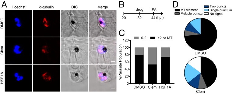
Fig. 3.
Clemastine but not HSF1A interferes with microtubule biogenesis during P. falciparum mitosis. (A) Representative confocal z-sections of P. falciparum-infected erythrocytes after treatment with 10 μM clemastine, 10 μM HSF1A, or 0.1% DMSO at 32 hpi for 12 h (n > 30). Cells were stained with Hoechst (blue) and anti–α-tubulin antibody (red) to visualize parasite nuclei and microtubule structures, respectively. Data are representative of two to three biological replicates. (B) Assay schematic showing drug administration and cell fixation for immunofluorescence assays (IFAs). (C) Parasites were scored as 0 to 2 tubulin puncta (0 to 2, gray bar) versus containing multiple tubulin puncta or microtubule filaments (>2 or MT, black bar), which showed clemastine treatment reduced late-stage morphologies by 32 ± 5.5% (mean ± SEM). Representative data of two to three biological replicates are shown. (D) Tubulin phenotypes were quantified for DMSO- and clemastine-treated parasites. Clemastine-treated parasites showed higher numbers with aberrant microtubule filaments or no tubulin signal when compared to the negative control. DIC, differential interference contrast. (Scale bar, 2 μm.)

