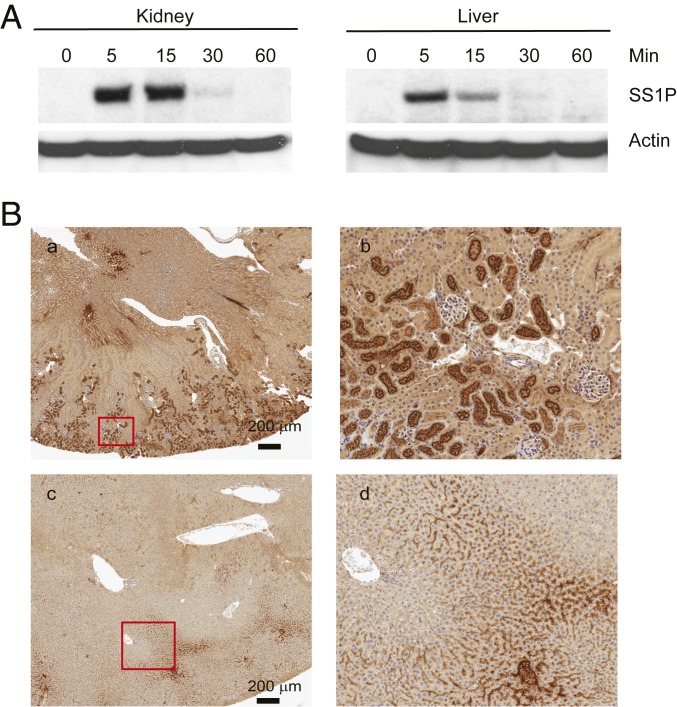
Fig. 2.
Immunotoxins in kidney and liver. (A) Western blot analysis of SS1P stability: Nude mice were injected with 30 μg of SS1P, and at indicated times, the kidney and liver were removed. Protein lysates were then analyzed by Western blot with anti-PE38. Actin was blotted as loading control. (B) Localization of SS1P in kidney and liver. Thirty micrograms of SS1P was injected in nude mice i.v., kidney (a and b) and liver (c and d) were removed at 15 min. The kidney and liver sections were immunohistochemically stained with anti-PE38. b and d are further magnification of the corresponding red box from a and c.

