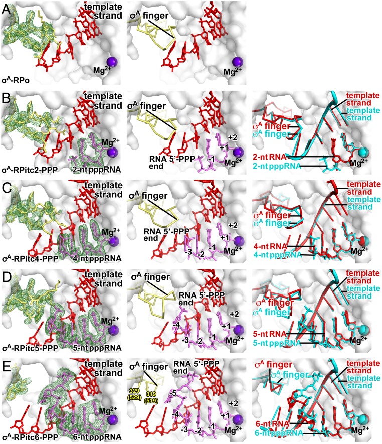
Fig. 2.
Stepwise, RNA-driven displacement of the σ-finger: primary σ-factor, 5′-triphosphate RNA. Crystal structures of (A) σA-RPo (PDB 4G7H; ref. 5), (B) σA-RPitc2-PPP (this work; see SI Appendix, Table S3), (C) σA-RPitc4-PPP (this work; see SI Appendix, Table S3), (D) σA-RPitc5-PPP (this work; see SI Appendix, Table S3), and (E) σA-RPitc6-PPP (this work; see SI Appendix, Table S3). Left subpanels, electron-density map and atomic model (colors and contours as in Fig. 1). Center subpanels, atomic model (colors as in Fig. 1). Right subpanel, superimposition of structures of corresponding complexes with 5′-hydroxyl RNA (cyan and gray) and 5′-triphosphate RNA (red and gray).

