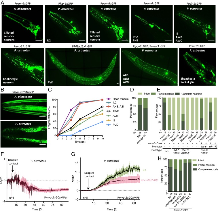
Fig. 5.
P. ostreatus triggers rapid cell necrosis via a mechanism that requires intact ciliated sensory neurons. (A) Images of cell necrosis in various neurons (indicated at the bottom left of each image) and glia cells. (Scale bars, 20 µm.) (B) Images of cell necrosis in the head or body wall muscle (Pmyo-3::mitoGFP). (Scale bars, 20 µm.) (C) Rates of cell fragmentation in various reporter lines that label specific neurons and head muscle (n > 20 for each time point). (D and E) Quantification of necrosis of ciliated sensory neurons (Posm-6::GFP) in wild-type N2 and ced-3 mutants (D), or Pleurotus-resistant mutants and cell-specific rescue lines expressing osm-6 cDNA under various promoters (E). (F) GCaMPer signal of the pharyngeal corpus of adult N2 in response to P. ostreatus hyphae (mean ± SEM; n shown above the x axis). (G) GCaMP6 signal of the pharyngeal corpus of adult N2 and unc-68(e540) mutants in response to P. ostreatus hyphae (mean ± SEM; n shown above the x axis). (H) Quantification of necrosis of ciliated sensory neurons in mutants in which calcium release is modulated from the ER.

