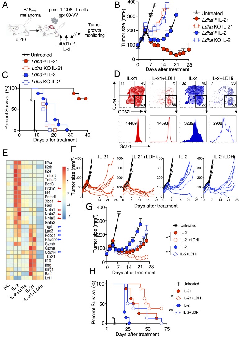
Fig. 4.
Inhibiting LDH in vitro enhances T cell priming and antitumor activity in vivo. (A) Schematic of the adoptive cell transfer protocol. pmel-1+ CD8+ T cells were cultured in the presence of anti-CD3 and anti-CD28 for 4 d, with concomitant stimulation with IL-2 or IL-21 in the absence or presence of LDHi. Cells were then adoptively transferred into animals that had been injected with B16 melanoma 10 d earlier. IL-2 was administered at the time of adoptive cell transfer and on each of the next 2 d. (B) Average tumor curves for transferred WT or Ldha KO CD8+ T cells treated with IL-2 or IL-21 or left untreated, as indicated. (C) Percent survival for each treatment group in B. (D) pmel-1 CD8 T cells were treated with IL-2 or IL-21 in the absence of presence of LDHi, and CD44 vs. CD62L expression was assessed on live cells (Upper). Gating was done on the CD62LhiCD44lo cells from the upper panels to assess Sca-1+ cells (histograms, Lower). (E) Heatmap of RNA-Seq data for cells treated with NC, IL-2, IL-2+LDHi, IL-21, or IL-21+LDHi. (F) Graphs showing individual tumor size over time for each mouse in the experiment. (G) Average tumor curves from data in F for untreated (black) or treated with IL-21 (solid red circles), IL-21+LDHi (open red circles), IL-2 (solid blue circles), or IL-2+LDHi (open blue circles). Statistics comparing tumor curves were performed using the Wilcoxon rank-sum test. (H) Percent survival for each treatment group in the tumor experiment shown in F and G. Statistics comparing percent survival data were performed using a log-rank (Mantel–Cox) test. All tumor data are from one of two similar experiments. *P < 0.05; **P < 0.01.

