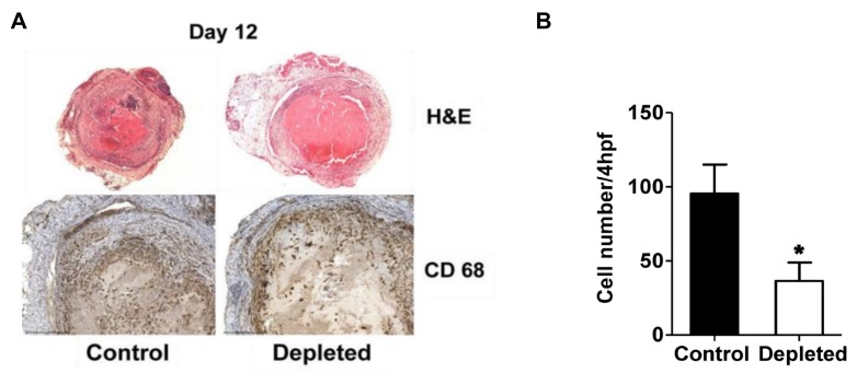
Figure 4.
Depletion of T cells reduces intra-thrombotic macrophage numbers. (A) Histochemical analysis by H&E staining of venous thrombi sections from control and T cell depleted mice at 12 days after vena cava ligation (upper panel) and immunohistochemical analysis of intra-thrombotic macrophage accumulation using anti-CD68 antibodies in venous thrombi sections from control and T cell depleted mice at 12 days after vena cava ligation (lower panel). Original magnification, ×100 upper panel, ×200 lower panel. Representative results from 4–5 independent animals are shown. (B) Quantification of the numbers of CD68 positive cells (macrophages). All values represent the mean ± SEM (n = 4–5). *p < 0.05, control versus T-cell depleted.

