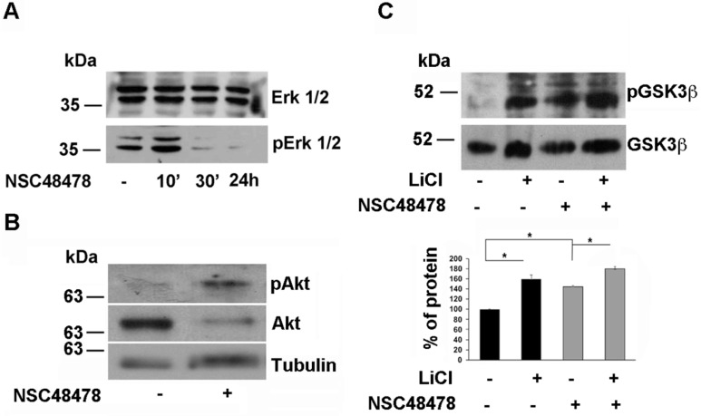
Figure 11.
NSC48478 induces inactivation of ERK1/2 axis and activation of Akt with consequent inactivation of GSK3β. (A) Total cell lysates (40 μg) from cells treated or not with NSC48478 for indicated times, were loaded on gels and expression levels of both total ERK1/2 and pERK1/2 were analysed by SDS-PAGE followed by Western blotting and hybridization of PVDF membranes by respective antibodies. (B) Total cell lysates (40 μg) from cells treated or not with NSC48478, were loaded on gels and expression levels of both total Akt and pAkt were analysed by SDS-PAGE followed by Western blotting and hybridization of PVDF membranes by respective antibodies. Anti-tubulin antibody was used to test loading controls. (C) The membranes were treated as in (B) with the exception that here the cells were incubated with LiCl (10 mM for 24 h) to inhibit GSK3β pathway, in the presence or absence of NSC48478. Both total and pGSK3β levels were revealed by using anti- GSK3β and anti-phospho-Ser9 GSK3β antibody, respectively. The amount of pGSK3β was quantified as “percent of protein” in the plot from three independent experiments (* p < 0.05).

