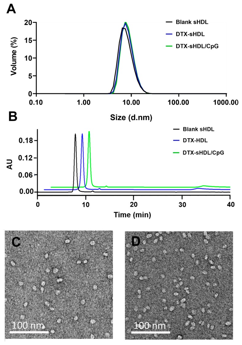
Figure 1.
Characterization of sHDL loaded with DTX and CpG. (A) Dynamic light scattering (DLS) analysis of blank sHDL (black curve), DTX-sHDL (blue curve), DTX-sHDL/CpG (green curve). (B) Gel permeation chromatography (GPC) analysis of blank sHDL (black curve), DTX-sHDL (blue curve), DTX-sHDL/CpG (green curve). (C,D) Transmission electron microscopy (TEM) images of DTX-sHDL (C) and DTX-sHDL/CpG (D) particles.

