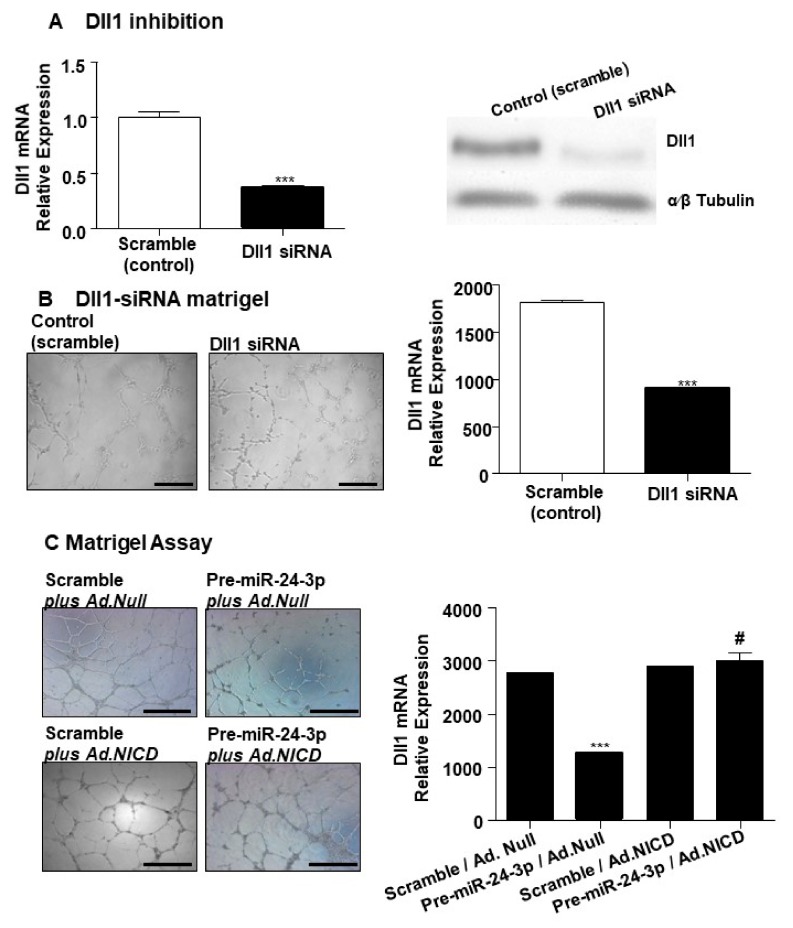
Figure 3.
Dll1 inhibition in HUVECs affects the network-formation capability, while Notch intracellular domain (NICD) over-expression rescues this angiogenic defect. (A, left) Bar graph shows Dll1 mRNA expression in HUVECs transfected with Dll1-siRNA (black column) compared to HUVECs transfected with scramble sequence (control). (A, right) Western Blot shows the Dll1 protein expression in HUVECs transfected with Dll1-siRNA compared to HUVECs transfected with scramble sequence (control). α-β Tubulin is presented as house-keeping protein. (B, left) Photomicrographs of representative fields show the endothelial networks formed by HUVECs transfected with Dll1-siRNA compared to control group. (B, right) Bar graph represents the quantification of the total length of cord-like structures formed by HUVECs transfected with Dll1-siRNA (black column) compared to control group (scramble, white column). (C, left) Photomicrographs of representative fields show the endothelial network formed by HUVECs differently treated as indicated: HUVECs transfected with scramble and infected with Ad.Null; transfected with pre-miR-24-3p miR-24-3p and infected with Ad.Null; transfected with scramble and infected with Ad.NICD; and HUVECs transfected with pre-miR-24-3p and infected with Ad.NICD. Bar graph (C) represents the photomicrographs’ conditions expressing the total length of tube-like structures. Dll1 mRNA expression was normalised to 18S. Experiments were performed in triplicate and repeated three times. Values are means ± SEM. *** p < 0.001 vs. Scramble in A nd B) and vs. Scramble/Ad.Null (in C); # p < 0.05 vs. Pre-miR-24-3p/Ad.Null (in C).

