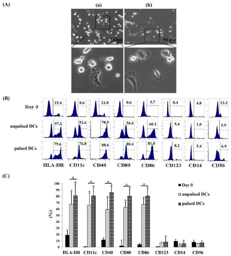
Figure 1.
Dendritic cells (DCs) differentiated from human cord blood (CB) monocyte cells. (A) Morphology of DCs differentiated from CB monocyte cells in the presence of granulocyte-macrophage colony-stimulating factor (GM-CSF), IL-4, and tumor necrosis factor (TNF)-α. (Aa) Morphology of DCs which were pulsed with A549 protein; (Ab) morphology of DCs which were not pulsed with A549 protein. The DCs exhibited many dendrites protruding from the cell bodies, which are characteristic of mature, pulsed DCs. (B) The percentages (%) of DC-surface markers expressed in pulsed DCs and unpulsed DCs. The pulsed DCs expressed high levels of dendritic surface markers including HLA-DR, CD11c, CD40, CD80, and CD86 while lower levels of these markers were detected on unpulsed DC. (C) The comparison of DC surface marker expression between unpulsed DCs and pulsed DCs. Noted that more than 80% of cells expressed DC markers in pulsed DCs population, meanwhile this value was more than 59.0% in unpulsed DCs. Data was presented as mean ± SD in quadruplicate cultures (* p < 0.05). pulsed DCs: A549 tumor cell lysate-pulsed DCs; unpulsed DCs: A549 tumor cell lysate-unpulsed DCs.

