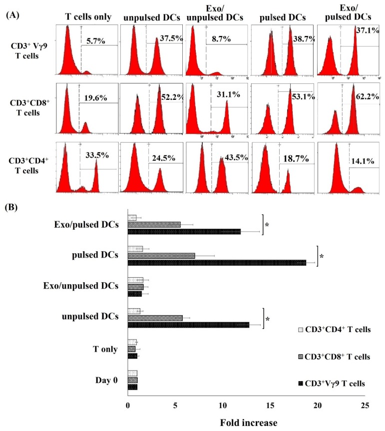
Figure 3.
DCs and their exosomes induced the proliferation of allogeneic T cell subpopulation. (A) After incubation with pulsed DCs, unpulsed DCs, and Exo/pulsed DCs, the percentage of both CD3+Vγ9 T cells and CD3+CD8+ T cells increased compared to T cells without such treatments. (B) Fold increase in the number of CD3+Vγ9 T cell and CD3+CD8+ T cell induced by DCs and their exosomes. Both pulsed DCs and their exosomes successfully induced CD3+Vγ9 T cell and CD3+CD8+ T cell proliferation, meanwhile the number of CD3+CD4+ T cells did not increase. Exo/pulsed DCs could induce the percentage of these cell sub populations similar to unpulsed DCs did, but exosomes isolated from unpulsed DCs did not. Data was presented as mean ± SD in quadruplicate cultures (* p < 0.05). DCs: dendritic cells; pulsed DCs: A549 tumor cell lysate-pulsed DCs; unpulsed DCs: A549 tumor cell lysate-unpulsed DCs; Exo/unpulsed DCs: exosomes isolated from unpulsed DCs; Exo/pulsed DCs: exosomes isolated from pulsed DCs.

