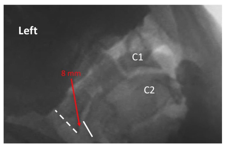
Figure 2.
Exemplar of AP open mouth left lateral flexion view of C1 on C2, demonstrating abnormal alignment. The dashed line indicates the lateral border of the left lateral mass of C1, and the solid line indicates the lateral border of the left articular pillar of C2. The red arrow indicates 8 mm lateral translation of C1 on C2 during maximal voluntary lateral flexion. Note: the image has been reversed so that left on the image corresponds with the patient’s left.

