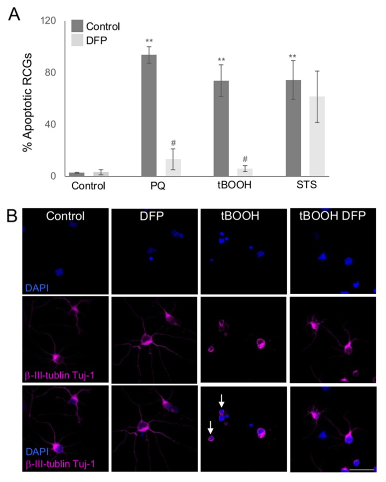
Figure 2.
DFP rescues RGC death from oxidative stress inducers. (A) Primary RGCs were incubated with 2 mM PQ, 200 µM tBOOH and 3.5 µM STS, in the presence or absence of 0.25 mM DFP for 24 h. After the incubation cells were fixed and stained with β-III-tubulin (TUJ1) and DAPI. The % of apoptotic RGCs was determined by counting TUJ1 positive cells with condensed apoptotic nuclei. The number of RGCs was counted in at least 5 fields of three different experiments. ** p < 0.01 vs. control; # p < 0.05 vs. treatment. (B) Representative images of cells treated as in A stained with β -III-tubulin (TUJ1, magenta) and nuclei (DAPI in blue). Examples of apoptotic nuclei are depicted with white arrows. Scale bar 50 µm.

