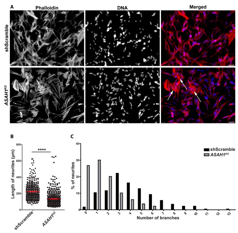
Figure 7.
ASAH1KD cells display short neurites and fewer branches per neurite. (A) Immunofluorescence microscopy images of differentiated shScramble and ASAH1KD cells stained with the phalloidin (Alexa Fluor® 568 dye, red). Nuclei were stained with Hoechst dye (blue). The scale bars correspond to 40 μm. White arrows show cells with stress fibers. (B) Length of neurites (μm) per cell. The mean neurite length was significantly lower in ASAH1KD cells compared to shScramble cells (**** p < 0.0001, Student t-test). (C) Distribution of the number of branches per neurite (p < 0.0001, Chi-square test).

