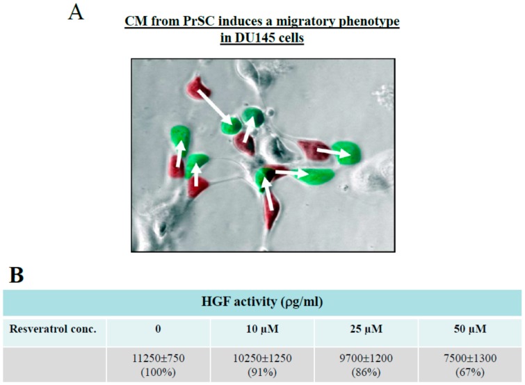
Figure 3.
(A) Time lapse microscopy analysis of migration of prostate DU145 cells, in response to CM from PrSC. Two images, one taken at time 0 h and a second one take at time 2.4 h, were overlaid using Adobe Photoshop. Cells at 0 h were marked red, and cells at 2.4 h were marked green. The coordinates for each cell were obtained for each of the two time points, and the change in the distance for each cell was shown. (B) Effect of treatment by resveratrol for 24 h on secreted hepatocyte growth factor (HGF) by PrSC, assayed by ELISA.

