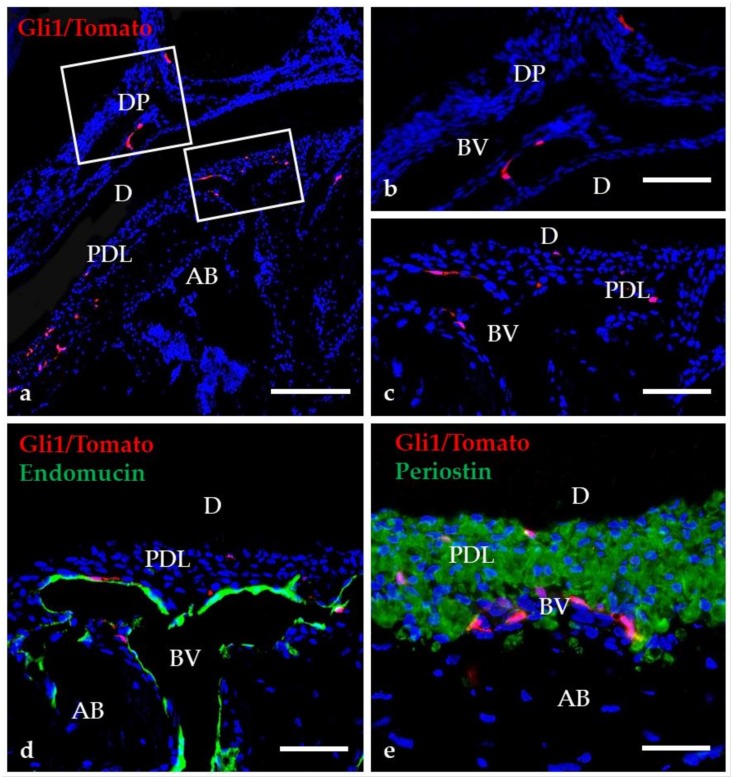
Figure 3.
Distribution of Gli1-positive cells in mature teeth. Higher magnification of the boxed region in “a” are shown in “b”–“e.” (a–c) Gli1-positive cells are present in the dental pulp (DP) and the periodontal ligament (PDL). (d–e) The merged image of Endomucin and Periostin with Gli1/Tomato fluorescence demonstrate that most Gli1/Tomato-positive cells are distributed near blood vessels (BV). AB, alveolar bone; D, dentin. Scale bars = 100 μm (a), 25 μm (b–e).

