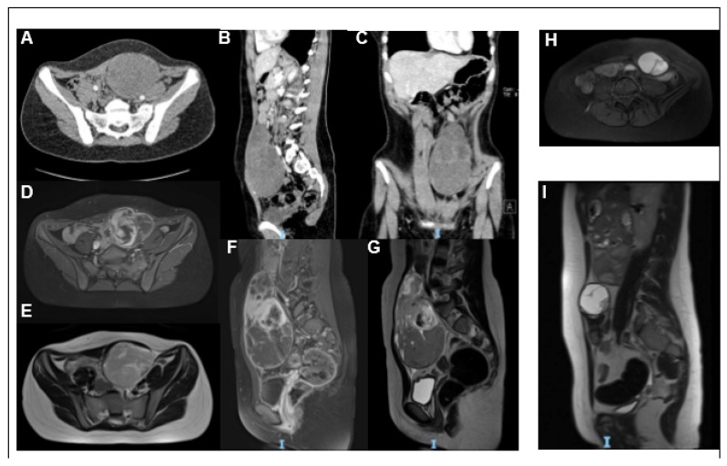
Figure 1.
Imaging: Axial (A), sagittal (B), and coronal (C) Computed tomography (CT) scan images showing a gross expansive non-infiltrating mass, with clear and well-defined margins and an oval and oblong appearance. In coronal sections, it clearly shows a bilobated aspect with a homogeneously hypodense apical portion and a well-capped lower portion, which has a more uneven density, as in fluid internal lacunae. The lesion has a maximum size of 81.9 mm × 77 mm and a deep cranium-caudal extension of approximately 14 cm, up to the pelvic floor and posterior to the lumbosacral spine, which appears to have a cleavage plane. Magnetic resonance imaging (MRI) images (D–I) of the expansive process at diagnosis (D–G) and after neoadjuvant chemotherapy (H–I). T1w-fat sat axial (D) and sagittal (F), T2w axial (E) and sagittal (G) images show a heterogeneous mass extending in the left abdominal wall, in the left paramedian seat at the level of the navel. T1w-fat sat axial images (H) and T2w sagittal (I) images show a clear reduction in the dimensions of the known expansive formation after chemotherapy.

