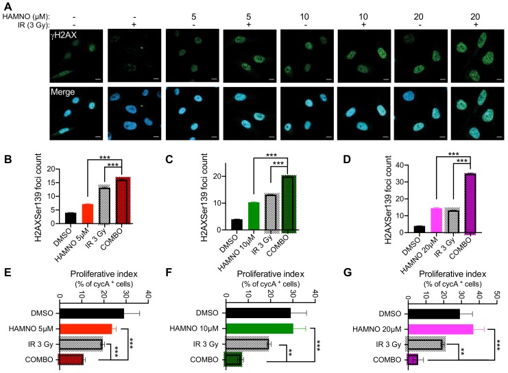
Figure 4.
HAMNO treatment induces DNA damage and activates cell cycle checkpoints in glioblastoma cancer stem-like cells. (A) Representative confocal microscopy images of γH2AX foci in G01-GSCs treated for 24 h with a vehicle control (DMSO) or increasing doses of HAMNO—5, 10 and 20 μM; then irradiated (COMBO) or sham-irradiated and incubated for an additional 24 h. (B–D) Microscopy-based quantification of γH2AX foci in G01-GSCs treated for 24 h with a vehicle control (DMSO) or increasing doses of HAMNO—5, 10 and 20 μM; then irradiated (COMBO) or sham-irradiated and incubated for additional 24 h. (E–G) Microscopy-based quantification of a proliferative index (% of nuclear cyclin A-positive cells) in G01-GSCs treated for 24 h with a vehicle control (DMSO) or increasing doses of HAMNO—5, 10 and 20 μM; then irradiated (COMBO) or sham-irradiated and incubated for additional 24 h. Data are presented as mean ± SD. Statistical significance was tested using one-way ANOVA, where ** p < 0.01, *** p < 0.001. N = 3.

