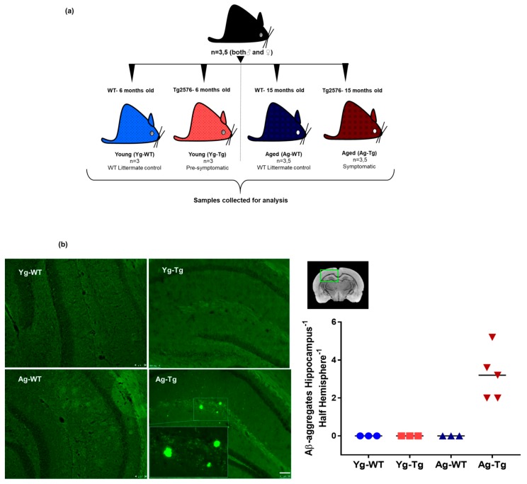
Figure 1.
Experimental design, timeline, and presence of cerebral Aβ pathology in Tg2576 mouse model. (a) Associated timeline in our animal model of pre-symptomatic at 6 months (Yg-Tg) and symptomatic at 15 months (Ag-Tg) timepoints. (b) Thioflavin S staining in Tg2576 mice for visualization of parenchymal Aβ plaques in coronal brain slices confirms the absence of Thioflavin S-positive plaques at 6 months and their presence in the subiculum and hippocampal formation at 15 months. n = 4 (or) 5. Data are expressed as mean + SEM, as well as individual values, and are obtained from >two independent experiments. Magnification 10X; section thickness- 15 μm. Scale bars: 100 μm WT wild-type, Tg-Transgenic; Green dots- Aβ plaques.

