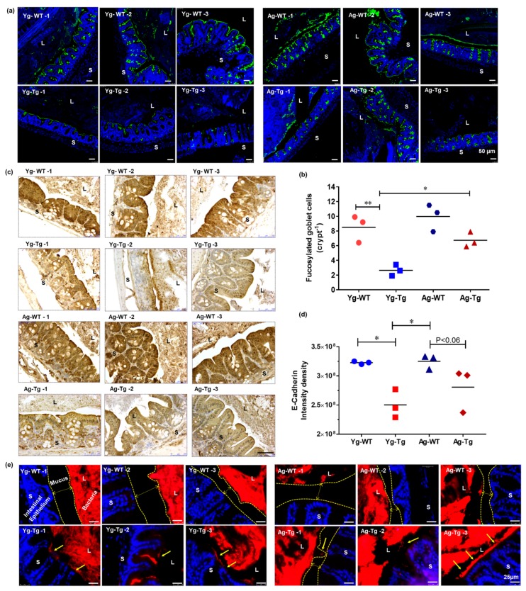
Figure 2.
Gut dysfunction occurs before development of Aβ pathology in the brain in Tg2576 mice. (a,b) Lectin staining and mucus fucosylation shows a significant reduction in mucus fucosylation with Ulex europaeus agglutinin staining of the terminal mucus fucose in the cecum of Tg2576 at 6 months when compared to age-matched WT controls. (c,d) Immunohistochemical staining of intestinal epithelial shows a significant reduction in E-cadherin expression in Tg2576 mice when compared to WT littermate controls at 6 months. (e) Widespread bacterial breach through the mucosal barrier and the corresponding antigenic load onto the intestinal epithelium detected by FISH in the cecum of Tg2576 mice at 6 months. n = 3 per group. Data are expressed as mean ± SEM, as well as individual values, and are obtained from >two independent experiments at various times. * p < 0.05, ** p < 0.01. P values were calculated using Two-Way ANOVA analysis with Tukey’s multiple comparisons test (b) and (d). Scale bars: 50 μm (a), 250 μm (c), 25 μm (e). WT wild-type, Tg-Transgenic. Green- mucus (a), brown- e cadherin (c) and red- bacteria (e), blue-DAPI nuclei.

