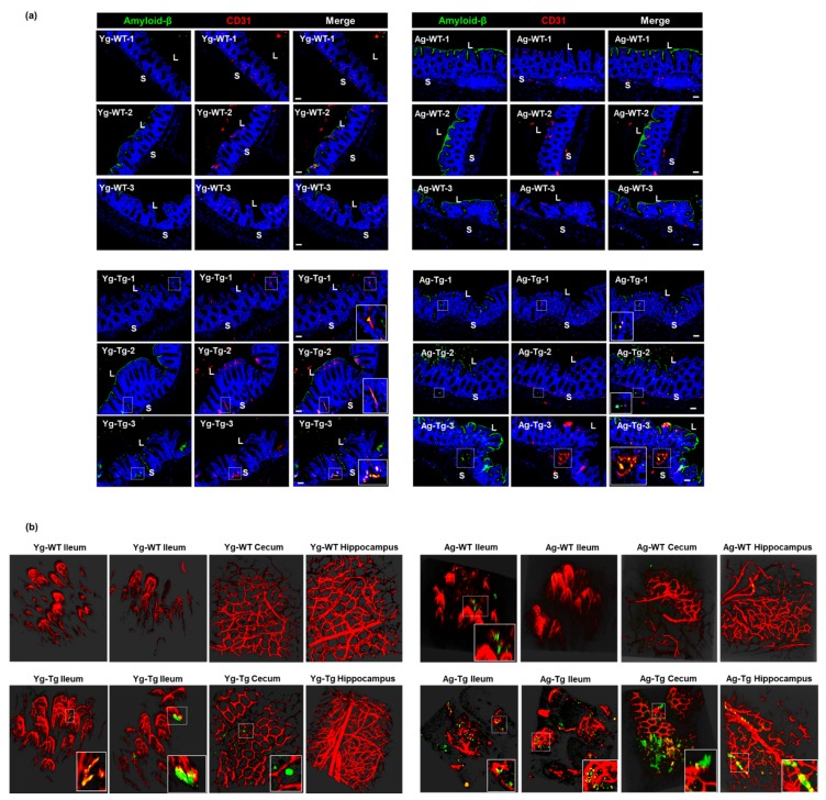
Figure 6.
Aβ co-localization with the intestinal vasculature in pre-symptomatic Tg2576 (Yg-Tg). (a) Immunohistochemical staining of Aβ by 4G8 antibody shows detectible Aβ deposits co-localized with vascular CD31 in the intestinal samples from Tg2576 at pre-symptomatic timepoint of 6 months (Yg-Tg) compared to age-matched WT littermate controls (Yg-WT) which significantly intensifies by 15 months (Ag-Tg). (b) Two-photon imaging of intestinal tissue shows detectible levels of Aβ accumulation with ileal and cecal vasculature in the gut but not in the hippocampus of the brain at 6 months in Tg2576 mice (Yg-Tg). Ileum, cecum and hippocampus samples show strong Aβ plaques co-localized with intestinal and cerebral vasculature at 15 months in Tg2576 mice (Ag-Tg). Data are obtained from >two independent experiments per group at various times (a) and separate set of two independent experiments at different times (b) due to dye injection. (a) Magnification 20×. Bar indicates 50 µm. L—luminal, S—serosal, Yg—young, Ag—aged, Tg—transgenic, WT—wild type. Red—CD31, Green—amyloid β, Blue—DAPI nuclei. (b) Red—vasculature, Green—amyloid β. (b) Magnification 20×.

