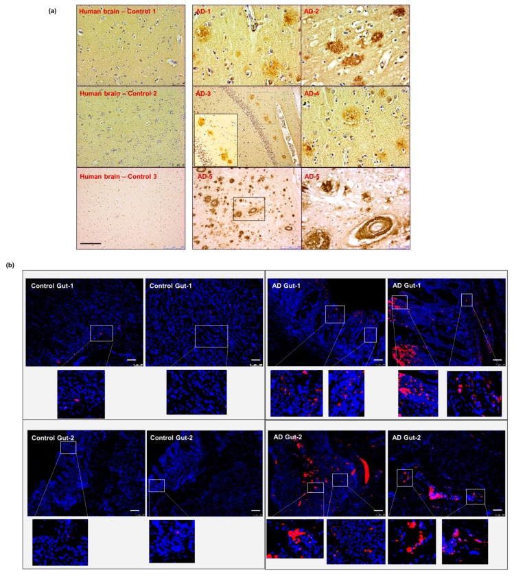
Figure 7.
Aβ detection in human sporadic AD gut and brain samples. (a) All five AD human brain samples show significant levels of parenchymal and perivascular Aβ depositions. (b) The gut tissues obtained from two of the same set of AD patients (corresponding to brain samples AD-1 and AD-2 in panel (a) show the presence of Aβ-aggregation on the epithelium and in the intestinal mucosa. Scale indicates 250 µm (a) and 100 µm (b). Magnification 20× (a). AD—Alzheimer Disease brain. Brown—amyloid β aggregates. (b) AD—Alzheimer Disease gut. Magnification 20× (b). Red—amyloid β, Blue—DAPI nuclei.

