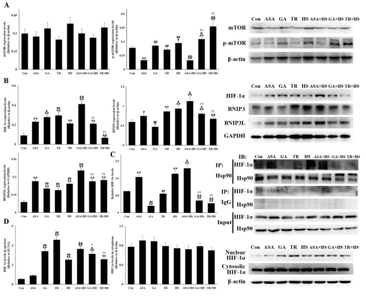
Figure 6.
The HIF-1α-BNIP3/BNIP3L pathway is involved in heat stress-induced autophagy. With the indicated antibodies, Western blot analysis was used to detect the levels of different proteins, and coimmunoprecipitation was used to detect the interaction of Hsp90 with HIF-1α. Data represent the means ± SD. (A) Total proteins of tested kidney tissues from all groups were analyzed for the levels of mTOR and p-mTOR by Western blotting. The relative abundance of the tested proteins was normalized to that of β-actin. (B) Total proteins of tested kidney tissues from all groups were used to detect the levels of HIF-1α, BNIP3, and BNIP3L by Western blotting. The relative abundance of the tested proteins was normalized to that of β-actin. (C) Total proteins of tested kidney tissues from all groups were subjected to coimmunoprecipitation to observe the interaction of Hsp90 with HIF-1α. The reactive bands from co-immunoprecipitation analysis were quantified using Image J software (Left). The abundances of bands in Con were set as 1. Representative bands are presented (Right). (D) Nuclear and cytoplasmic proteins were extracted to detect HIF-1α levels. The relative abundance of the tested proteins was normalized to that of PCNA (band not shown) and β-actin. The comparison between the Con group and other groups is indicated by * p < 0.05 and ** p < 0.01, comparison of the ASA group with the GA, TR, and HS groups is indicated by # P < 0.05 and ## p < 0.01, comparison of the HS group with the ASA+HS, GA+HS, and TR+HS groups is indicated by & p < 0.05 and && p < 0.01, and comparison of the ASA+HS group with the GA+HS and TR+HS groups is indicated by † p < 0.05 and †† p < 0.01.

