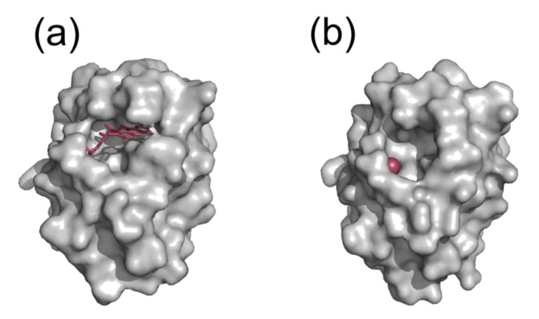
Figure 3.
Molecular surface representation of holo (a) and apo (b) flavodoxin from Hp. FMN cofactor and a chloride ion bound at the FMN phosphate site are shown as red sticks and a sphere, respectively. The two structures are similar and exhibit an unusual pocket close to the cofactor binding site. Most other (apo)flavodoxins lack such surface pocket.

