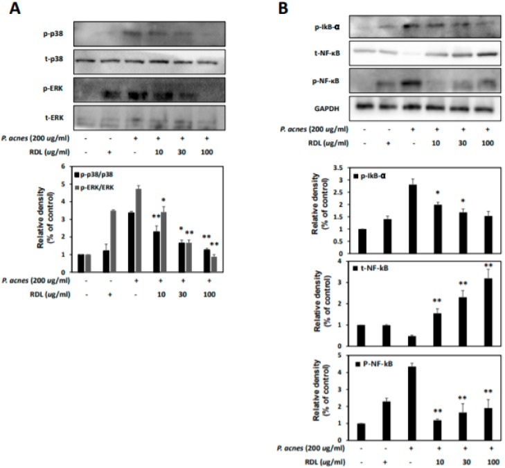
Figure 4.
RDL suppresses the MAPK and NF-κB signaling pathway in P. acnes–treated HaCaT cells. HaCaT cells were incubated for 24 h without P. acnes and with P. acnes alone, or with P. acnes in the presence of RDL. (A) The effects of RDL on MAPKs, such as P38 and ERK phosphorylation, in HaCaT cells. P38 and ERK were quantified with ImageJ. (B) Effects of RDL on phosphorylation of IkB and NF-κB after P. acnes treatment of HaCaT cells. Western Blots were quantified using densitometry and normalized to total protein and GAPDH. p-IκB, NF-κB and p-NF-κB were quantified with ImageJ. Data were expressed as the mean ± SD three independent experiments. * p <0.05 and ** p <0.01 compared to the P. acnes only treatment.

