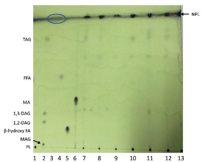
Figure 1.
Thin-layer chromatography (TLC) plate separation of bacterial lipids extracted from whole cells using chloroform:methanol (unbound MA extract), developed with the H:D:F 70:30:0.2 solvent system. Lanes correspond to: 1. PL; 2. MAG, DAG, TAG (Supelco lipid standard 1787-1AMP); 3. HC (blue circle at the top of the plate shows the diffuse area where this standard typically ran); 4. FFA; 5. β-hydroxy FA; 6. MA (mycobacterial free MA standard); 7. Williamsia sp. 1135; 8. Williamsia sp. 1138; 9. Rhodococcus qingshengii 1139; 10. Rhodococcus erythropolis 1159; 11. Rhodococcus sp. 1163; 12. Rhodococcus sp. 1168; 13. C. glutamicum. NPL = Non-polar lipid spots in bacterial extracts running with the solvent front at the top of the plate.

