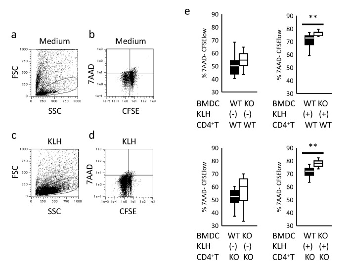
Figure 4.
Antigen presentation by BMDC and antigen-immunized CD4+ T-cells proliferation. CD4+ T-cells were isolated from WT and KO mice immunized with keyhole-limpet hemocyanin (KLH). CD4+ T-cells were co-cultured with KLH-stimulated BMDCs, as described in Materials and Methods. (a) Gating of CD4+ T-cells cultured with unstimulated KO BMDCs. (b) Proliferation of CD4+ T-cells cultured with unstimulated KO BMDCs. (c) Gating of CD4+ T-cells cultured with KLH-stimulated KO BMDCs. (d) Proliferation of CD4+ T-cells cultured with KLH-stimulated KO BMDCs. (e) Proliferation of CD4+ T-cells isolated from KLH-immunized WT and KO mice and co-cultured with unstimulated and KLH-stimulated BMDCs from WT and KO mice. Data are collected the data of two independent experiments each involving eight mice (WT n = 4, KO n = 4) analyzed by Student’s t test. ** p < 0.01.

