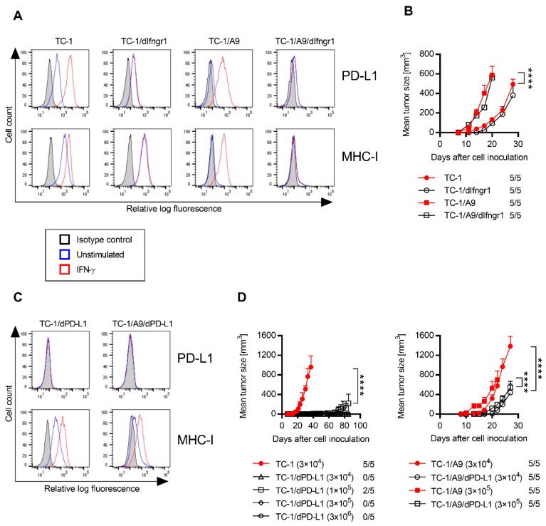
Figure 1.
Characterization of the derived cell lines. Surface programmed cell death protein 1 (PD-1) ligand 1 (PD-L1) and major histocompatibility complex class I (MHC-I) expression on unstimulated and stimulated (200 IU/mL interferon (IFN)-γ for 1 day) cells were analyzed by flow cytometry in TC-1, TC-1 clone with a deactivated IFN-γ receptor 1 (IFNGR1; TC-1/dIfngr1), TC-1/A9, and TC-1/A9/dIfngr1 cell lines (A) and TC-1/dPD-L1 and TC-1/A9/dPD-L1 cell lines (C). Cells were incubated with specific antibodies or isotype control antibodies. (B) Oncogenicity of TC-1, TC-1/dIfngr1, TC-1/A9, and TC-1/A9/dIfngr1 cell lines was compared after subcutaneous (s.c.) administration of 3 × 10⁴ cells to C57BL/6 mice (n = 5). (D) For the evaluation of oncogenicity of cell lines with deactivated PD-L1, various cell doses were s.c. injected. The ratio of mice with a tumor to the total number of mice in the group is shown. Bars ± SEM; **** p < 0.0001.

