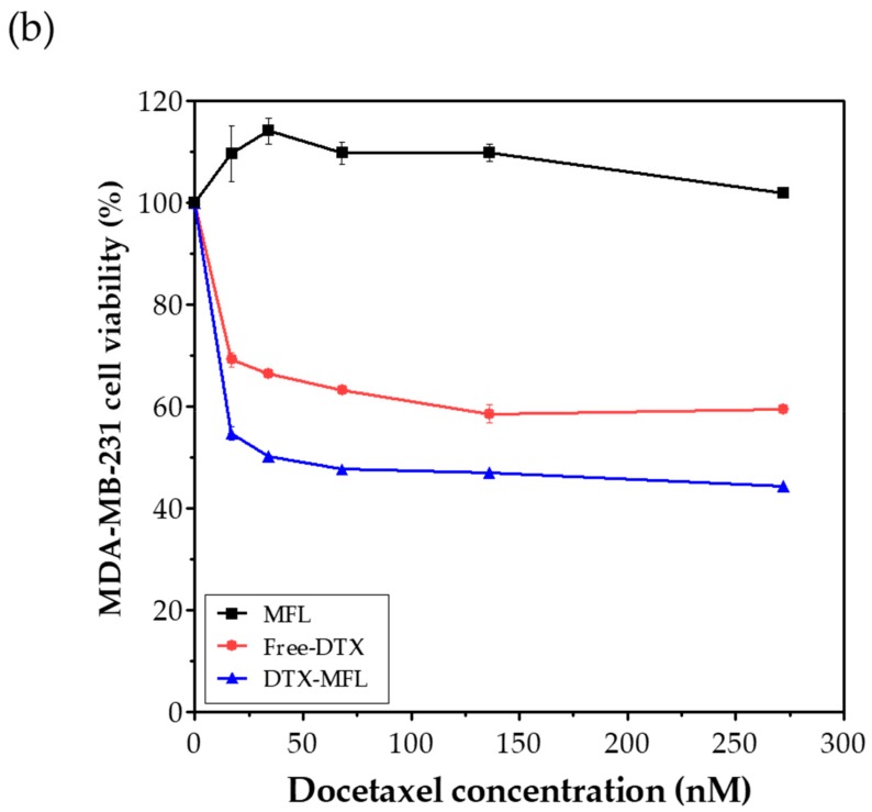
Figure 3.
(a) Confocal fluorescent microscopic images of MDA-MB-231 cells treated with membrane fusogenic liposomes and non-fusogenic liposomes loaded with fluorescent dye DiI. (b) Cell viability of MFLs, free-DTX, and DTX-MFLs at 24 h. MFL and NFL denote membrane fusogenic liposomes and non-fusogenic liposomes, respectively. Nuclei were stained with Hoechst (blue). Scale bars represent 20 μm.


