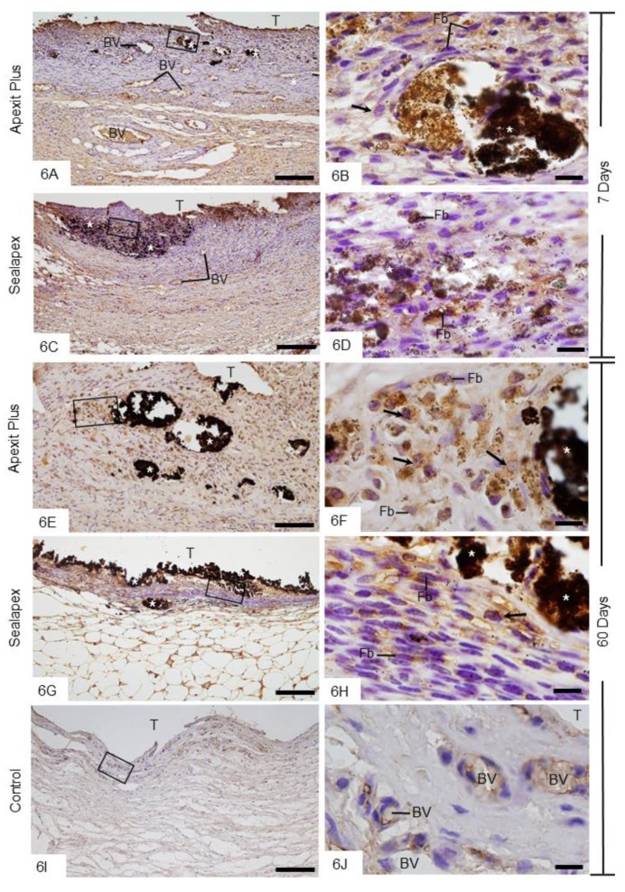
Figure 6.
Photomicrographs show sections submitted to the von Kossa method (black) followed by immunohistochemistry for detection of ALP (brown-yellow) and counterstained with hematoxylin. A,C,E,G General view of the capsules showing von Kossa-positive structures (asterisks). Note that ALP-immunolabelled cells are located mainly next to the von Kossa-positive structures (asterisks) and in the internal surface of capsules. B,D,F,H Higher magnifications showing outlined area of the general view. Fibroblasts (Fb) and round/ovoid cells (arrows) show strongly ALP-immunolabelled cytoplasm (brown-yellow). I,J None or weak ALP immunolabelling is present in the capsule of control group. In Figure 6J, outlined area of Figure 6I, some vascular cells exhibit subtle ALP immunoreactivity. T, space of the implanted tube; BV, blood vessel profiles. Bars: 150 µm (A,C,E,G,I) and 10 µm (B,D,F,H,J).

