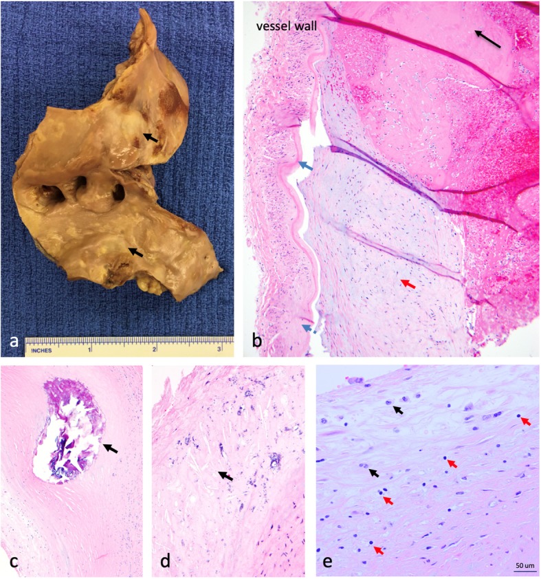
Fig. 2.

Gross and microscopic pathology. a Gross pathology of aortic arch revealing extensive atheromas (black arrows). b Histopathology of the anterior cerebral artery. There is intimal thickening underlying the internal elastic lamina (blue arrow); the media and adventitia are distorted by fibrosis (blue dashed arrow). A recently-formed fibrin thrombus (black arrow) is adherent to a chronic and organized atherosclerotic plaque (red arrow). c Cross-sectional view of the left anterior descending coronary artery containing an intramural calcified nodule (black arrow). d Cross-sectional view of the basilar artery wall, demonstrating architectural distortion with cholesterol clefts (black arrow) and microcalcifications. e Left anterior descending artery with thickening of the intima and inflammatory infiltrate including T lymphocytes (red arrows) and macrophages (black arrows)
