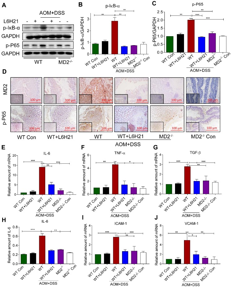
Figure 6.
MD2 blockade inhibits NF-κB activation in AOM/DSS mouse model. WT or MD2-/- mice were treated with AOM/DSS to produce a colitis-associated colon acncer growth (Methods). WT mice were also orally administered L6H21 (60 mg/ml) or 1% CMC-Na (vehicle control); n=10. (A) Western blot analysis of phosphorylated IκB-α and phosphorylated NF-κB P65 subunit (p-P65) in mouse colon tissues; GAPDH used as loading control. The immune-reactive bands were quantified by densitometry using Image J analysis: (B) p-IκB-α and (C) p-P65; values normalized to GAPDH. (D) Immunohistochemical staining of colon tissues from mice for MD2 (upper row; brown) and phosphorylated p65 subunit of NF-κB (bottom row; brown); n=6. Quantification (data in D) of MD2 intensity and phosphorylated NF-κB p65 subunit are provided in supplementary file. Real-time qPCR determination of pro-inflammatory genes in colonic tissue; mRNA values normalized to β-actin and reported relative to WT Con: (E) IL-6, (F) TNF-α, (G) TGF-β; n=3. (H) ELISA detection of IL-6 in colon tissue lysates; values normalized to total protein; n=6. Real-time qPCR determination of adhesion molecules in colonic tissue; mRNA values normalized to β-actin and reported relative to WT Con: (I) ICAM-1, (J) VCAM-1; n=3. Data in B, C, E, F, G, H, I, J are shown as mean±SEM; *P<0.05, **P<0.01, ***P<0.001.

