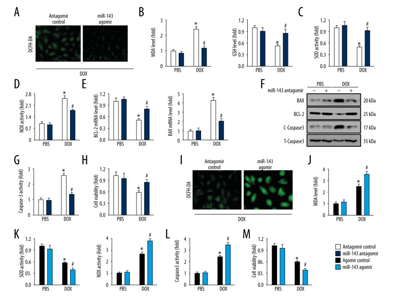
Figure 4.
MicroRNA-143 (miR-143) regulated oxidative stress and myocardial apoptosis in response to doxorubicin in vitro. (A) Representative images of dichloro-dihydro-fluorescein diacetate (DCFH-DA) staining of H9C2 cells (n=6). (B) Malondialdehyde (MDA) and glutathione (GSH) levels in cultured H9C2 cells (n=6). (C, D) Superoxide dismutase (SOD) and NADPH oxidase (NOX) activities in cultured H9C2 cells (n=6). (E) The relative mRNA levels about BCL-2 and BAX in cells (n=8). (F) Representative images of immunoblots (n=6). (G) Data on caspase-3 activity in cells (n=6). (H) Cell viability detected using the cell counting kit-8 (CCK-8) assay (n=6). (I) Representative images of DCFH-DA staining (n=6). (J) MDA production in cultured H9C2 cells (n=6). (K) SOD and NOX activities in H9C2 cells (n=6). (L) Quantitative data on caspase-3 activity in H9C2 cells (n=6). (M) Statistical analysis of cell viability in vitro (n=6). Data are presented as the mean±standard deviation (SD) with the 95% confidence interval (CI). * P<0.05 versus the normal saline (NS)+antagomir control group. # P<0.05 versus the doxorubicin+antagomir control group.

