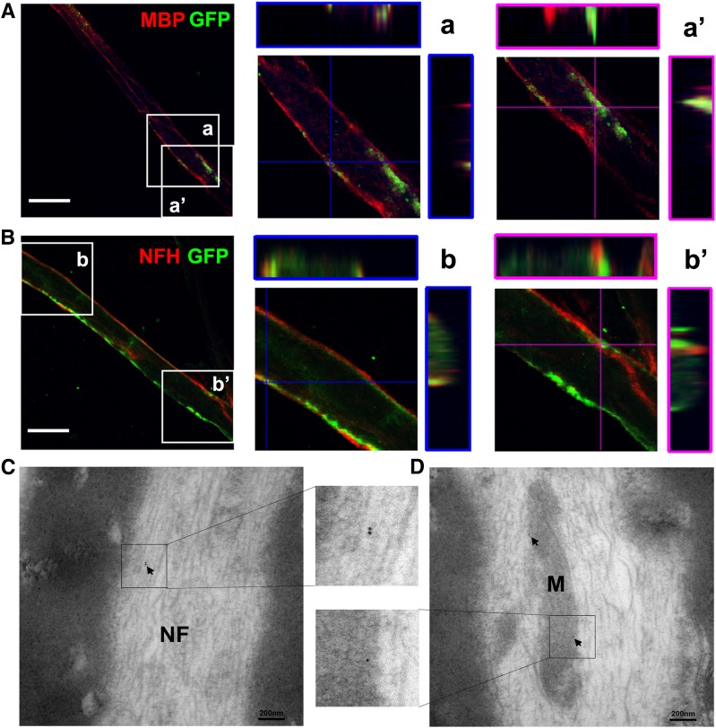
Figure 4.
Distribution of SC-Exos in the sciatic nerve tissue. Representative confocal microscopic images show that these nerve fibers are MBP positive (red) (A) and NFH positive (red) (B) with the presence of puncta GFP (green) signals. The orthographic three-dimensional projection in enlarged areas demonstrated that GFP signals are colocalized to MBP (a) and NFH (b). However, some of GFP signals are localized within the MBP-positive signals (a’) and outside the NFH-positive signals (b’). These data suggest that SC-Exos-GFP are internalized by MBP-positive Schwann cells and NFH-positive nerve fibers of the sciatic nerve. TEM images show that GFP immunogold-positive particles (arrows) are present in the neurofilament (NF) (C) and mitochondria (M) (D) of sciatic nerves, indicating the presence of SC-Exos-GFP in nerve fibers of the sciatic nerve. n = 3 mice/group. Scale bars = 20 μm (A and B).

