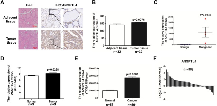
Figure 1.
ANGPTL4 is highly expressed in thyroid cancer compared with adjacent normal thyroid tissue. (A) Representative images of ANGPTL4 immunohistochemical staining in thyroid cancer and its adjacent tissue. Scale bars, 200 μm and 50 μm. H&E, hematoxylin and eosin. (B) ANGPTL4 protein levels in 32 human thyroid cancer and its adjacent tissue were analyzed by immunohistochemistry, p<0.0001. (C) ANGPTL4level increased in thyroid cancer compared with begin thyroid tissue according to RNA-Seq. (D) ANGPTL4 expression level was up-regulated in thyroid cancer tissues compared with matched adjacent normal tissues in GEO dataset. (E) ANGPTL4 expression in thyroid cancer (N = 501) and normal thyroid tissues (N = 58), p<0.0001. Data were obtained from TCGA dataset. (F) ANGPTL4 expression level was upregulated in thyroid cancer tissues compared with matched adjacent normal tissues in TCGA dataset.

