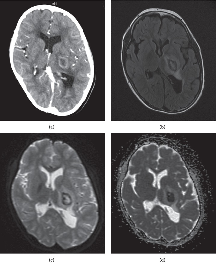
Figure 1.
Brain abscess detected by contrast-enhanced computed tomography and magnetic resonance imaging. (a) Contrast-enhanced computed tomography scan showing a low-density area with ring enhancement. (b) T2-weighted fluid-attenuated inversion recovery image showing a hyperintense lesion surrounded by a thin hypointense ring. Hyperintense edema is noted. (c) Diffusion-weighted magnetic resonance image demonstrating a hyperintense signal within the lesion. (d) Apparent diffusion coefficient map shows hypo-intensity within the lesion, confirming the presence of restricted diffusion.

