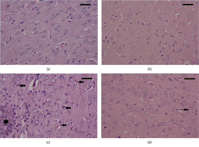
Figure 9.
Histopathological observations in cortical tissue following the treatment with red beetroot extract (RBR) and chlorpyrifos (CPF). (a) Photomicrograph of the cortical tissue of the control group showing healthy cortical structure. (b) Photomicrograph of the cortical tissue of rats treated with RBR alone showing a healthy histological structure. (c) Photomicrograph of the cortical tissue of rats exposed to CPF showing degenerative alterations in neurons, and a large number of eosinophil leukocyte infiltrating (black star) between neurons and apoptotic cells (arrow) have been observed. (d) Photomicrograph of the cortical tissue of rats treated with RBR and CPF showing a recovery of cortical tissue; however, some neurons are showing a degree of degeneration with infiltrating eosinophil. Sections were stained with hematoxylin and eosin (400x). Scale bar = 50.

