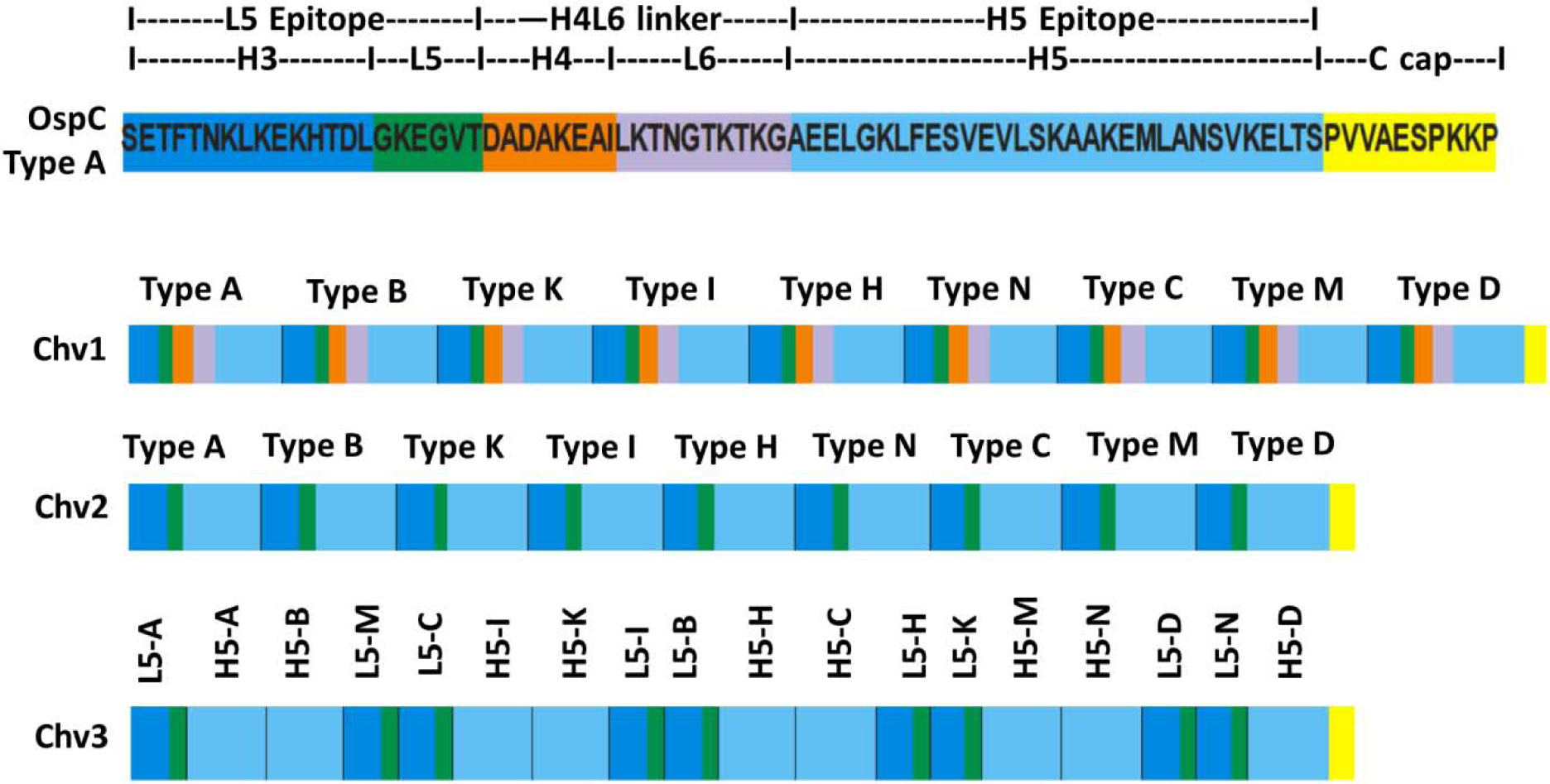
Figure 1. Schematic depiction of the organization of the Chv series of chimeritopes.

The top schematic is a linear representation of the Loop 5 (L5) and Helix 5 (H5) region of a type A OspC (B. burgdorferi B31) with the intervening sequence (Helix 4 Loop 6; H4L6) and structural elements shown. Coloring is used to highlight each structural domain. The lower schematics depict the organization of the Chv1, Chv2 and Chv3 chimeritopes. The color coding is the same as in the top panel and is included to highlight the organizational differences of each chimeritope.
