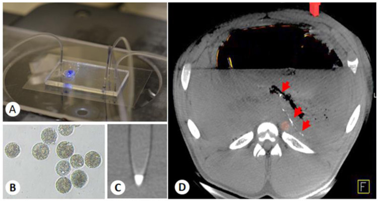
Figure 1.
(A) Custom-made microfluidic device for x-ray-visible, small-size, monodisperse, embolic microsphere synthesis. (B) Microscopic image of microfluidic-prepared, barium sulfate-impregnated alginate microspheres. (C) Maximum-intensity projection of a barium alginate microsphere phantom acquired with a clinical x-ray flat panel angiographic system (Axiom Artis Zee), demonstrating the radiopacity of the microspheres. (D) Axial view of the CBCT image of a pig stomach embolized with 50 μm barium alginate microspheres, showing the distribution of the microspheres one week after embolization (arrows).

