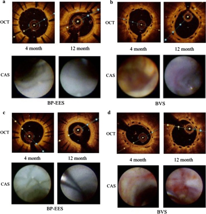
Fig. 2.
Representative OCT and CAS image at 4 and 12 months. The OCT images in upper panel, and CAS images in lower panel. a, c Follow-up images at 4 and 12 months after BP-EES implantation. OCT images showed almost all struts of BP-EES were covered at 4 months although the thickness of neointima was thin. CAS images of BP-EES demonstrate white homogenous neointimal coverage (NIC) grade 1–2 at 4 months. A small red intra-mural thrombi (arrow) at proximal edge in a. At 12 months, NIC grade was 3, without thrombus in both cases. b, d follow-up images at 4 and 12 months after BVS implantation. OCT images showed most scaffolds were well covered even at 4 months. At 12 months, the neointimal thickness did not increased. CAS images of BVS revealed severe ELI (erosion-like intima) at 4 months in both cases. At 12 months, ELI was attenuated but still observed. OCT optical coherence tomography, CAS coronary angioscopy

