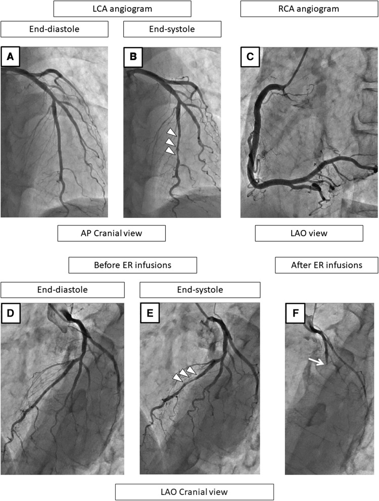
Fig. 2.
A representative case with MB and SPT-positivity in the LAD on the coronary angiogram at baseline (a–c), before (d, e), and after the SPT (f). At baseline and before the ER injections, the LCA angiogram shows no stenosis during the end-diastole phase (a, d) but MB is observed in the mid-LAD, as shown by squeezing (arrow heads) during the end-systole phase (b, e). The RCA angiogram shows no organic stenosis (c). After the ER injections, the LAD becomes a total occlusion at the proximal lesion (f). After the ER injections into the LCA, the chest pain and ECG changes persist and nitrates are required to relieve the vasospasms, and therefore, SPT in the RCA was not performed. In this case, the culprit vessel of the SPT-positivity was the LAD, and the positional relationship of the vasospasms to the MB was the “segment proximal to the MB” (f). CAG coronary angiography, SPT spasm provocation test, ER ergonovine, LAD left anterior descending coronary artery, RCA right coronary artery, ECG electrocardiogram

